Histology and Imaging Core (HIC)
Purpose
Research histopathology service
Description
To provide researchers fee-for-service access to expertise and state of the art instrumentation across a broad range of services including; routine histology, immunohistochemistry, microscopy, digital imaging and quantitative digital pathology, cell based multiplex assays and comparative pathology consultation.
This core is open to all investigators, including; UW and their affiliates, commercial and not-for-profit biotechnology companies, and other academic institutions.
Type: Equipment/Services
Services
- Routine histology including tissue processing and embedding (paraffin and OCT cryopreservation).
- Slide cutting and staining (H&E as well as affinity based histochemical (special stains).
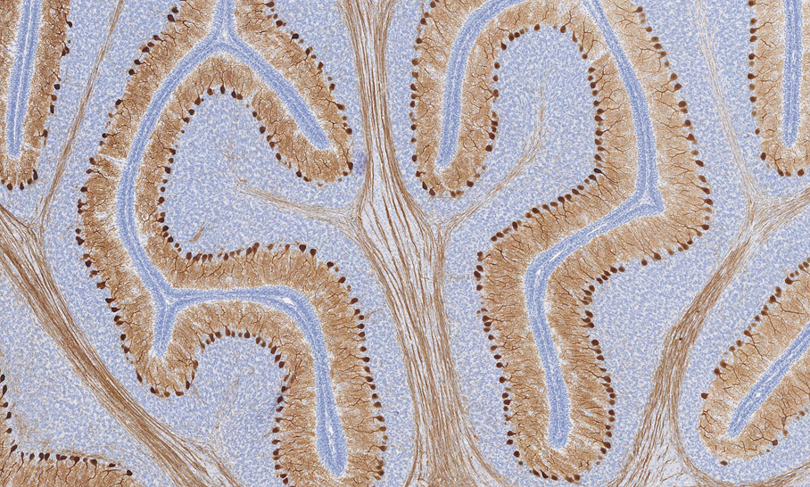
- Immunohistochemistry, including antibody optimization and validation as well as consultation on staining strategy and antibody selection.
- Whole slide scanning, image analysis and stereology
- Conventional upright brightfield and fluorescent microscopy and image acquisition
- Luminex Cell Based Multiplex Assay
- Comparative Pathology Consultation from board certified Veterinary Pathologists
Equipment
- Leica Bond
- Hamamatsu Nanozoomer Digital Pathology
- Visiopharm Integrator Software
- Nikon 90i upright research microscope
- Luminex Bio-Plex 200
- Deltavision Elite microscope
Keywords: General
Histology, Immunohistochemistry, Immunofluorescence, Digital Imaging, Quantitative Digital Pathology
Keywords: Specific
Histopathology, fixation, tissue processing, paraffin embedding, paraffin sectioning, frozen sections, cyrosections, IHC, antibodies, multiplexing, whole slide scanning, image analysis, stereology, Leica Bond, Visiopharm, Nanozoomer