February 16, 2006
New PET/CT scanner a national first
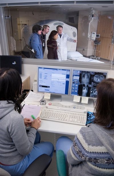
UW Medical Center is the first hospital in the country to install a new-generation PET/CT imaging system designed to help physicians detect, diagnose and monitor treatment of cancer and other diseases, including heart disease and neurological disease, more accurately and earlier in the disease process.
PET (positron emission tomography) and CT (computed tomography) scans are both standard imaging tools that physicians use to pinpoint disease states in the body. A PET scan demonstrates the biological function of the body before anatomical changes take place, while the CT scan provides information about the body’s anatomy such as size, shape and location.
The new-generation PET/CT is a fusion of the high-speed, high-resolution capabilities of a CT scanner with the metabolic and physiologic capabilities of a PET scanner, said Dr. Paul Kinahan, associate professor of radiology and director of PET/CT physics for UW Medical Center.
“By combining these two scanning technologies, a PET/CT scan enables physicians to more accurately diagnose and identify cancer, heart disease and brain disorders,” said Dr. Satoshi Minoshima, professor and vice-chair for research at the UW Department of Radiology.
“A PET/CT image allows physicians to see cancer earlier, localize and personalize treatment and carefully monitor that treatment,” said Dr. Hubert Vesselle, associate professor of radiology.
“It is becoming the tool of choice for oncology applications today and we’re excited to see how it can transform other clinical specialties.
“As data demonstrating the advantages of PET/CT over MRI and CT in early disease detection continue to accumulate, it is reasonable to anticipate that PET/CT imaging systems could become the first line of defense in optimization of treatment for cancer patients.”
While PET/CT is most commonly used for cancer diagnosis and treatment planning, its use is expanding into other areas, including cardiovascular and neurological imaging.
For cardiovascular imaging, the PET/CT scanner allows physicians to access essential metabolic and anatomical data including mapping of blood supply to heart muscle, CT angiography and cardiac calcium score.
The information made available through the combination system can help physicians accurately diagnose cardiac patients and help eliminate unnecessary invasive procedures.
In neurology, clinicians have been making strides in earlier detection of neurological diseases using PET/CT technology.
“PET/CT is a great diagnostic tool and powerful enough to accurately and quickly image brain metabolic abnormalities associated with neurological disorders such as Alzheimer’s disease,” Minoshima said.
In 2005, UW Medical Center installed the first hospital-based 64-slice CT scanner in the country. The Department of Radiology is currently installing the most advanced MR imaging system at South Lake Union and at UWMC.
“It nicely demonstrates how the excellence of our clinicians and researchers positions us to provide the best in care today and ensures a continued impact into the future,” said Dr. Norman Beauchamp, chair of the UW Department of Radiology.


