December 1, 2005
Seeking to understand adult stem cells
Less than a decade ago, researchers were able to confirm the existence of adult stem cells hiding out in the central nervous system, and ever since people have been trying to figure out how these cells could be activated to repair spinal cord injuries or even to cure neurodegenerative diseases.
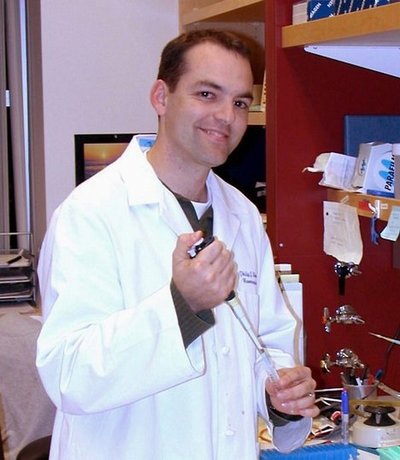
At the UW, Dr. Philip Horner’s laboratory has focused on understanding these adult stem cells and trying to learn how to manipulate them and the environments in which they grow.
“In the first place,” Horner says, “it’s important to know that these adult stem cells are quite different from the embryonic stem cells other labs are working with. The embryonic stem cells have the potential to become all kinds of tissues and structures, so the problem with them is learning how to establish enough control to make them become what you want them to be. With the adult stem cells, the problem is how to activate them.”
One advantage of working with the adult cells is that, more or less obviously, they are already there, not only in the body but in the parts of the body that need neurons replaced or repaired. Avoiding the need to bring the cells into the body could be a great advantage. And adult stem cells are naturally limited in their growth possibilities — they can only produce cells for the type of tissue in which they are found.
But so far no one has been able to understand exactly what needs to be done to cause the adult stem cells to go to work, grow new neurons and repair a severed spinal cord.
Researchers do know that in some areas of the brain, especially areas devoted to learning and memory, new neurons are being formed from adult stem cells. And some lower vertebrates, including salamanders, do have the ability to repair or replace some of their spinal cords, although no mammals can do it. Study of the successful repairs in salamanders shows that the repairs do begin with an activation of existing stem cells, Horner says.
The observations about adult stem cells growing neurons in some parts of the brain, but not in others, have led to careful examination of the local environments in which the stem cells are found. Each stem cell originates in a specific local environment neuroscientists call a “niche,” and that niche powerfully determines the cell’s fate. “In a way,” Horner said, “a niche is like the neighborhood in which a person grows up. It has long-lasting effects on what happens later and what the possibilities are.”
“For a long time, neuroscientists were just focusing on neurons themselves,” he explained. “But there’s been a real shift in thinking over the past few years, and we now believe that many of our answers may lie in the glial cells surrounding the neurons. We’ve always known they were there, but they used to be seen as just supporting tissue. Many recent findings point to the glial cells as controlling and regulating the development of neurons.”
One of the things these glial cells in the spinal cord may be doing is keeping the adult stem cells in a sort of “hibernating” state.
When one of the stem cells is removed and put in a growth medium with the right technique, Horner says, it will readily grow into a new neuron. But put it back in the original position, and it shuts down.
So the question may be not so much what can be given to the cells to encourage them, but what controls need to be found and removed.
To examine the stem cells, glial cells and neurons involved in these studies, Horner’s lab uses a variety of techniques, including genetic manipulation. A recent development allows them to prepare a piece of brain tissue so that they are able to see images, over a timespan of about a month, of a developing stem cell and how it interacts with other cells in its environment. Some of these images are made using a green fluorescent protein, so that they can track any cells that derive from the original stem cell.
Horner will describe the work of his lab in the next Science in Medicine Lecture at noon on Thursday, Dec. 8, in T-625 of the Health Sciences Center. It will be simulcast to the VA medical center in room 518, Building 1, and to Harborview in room 121 of the Research and Training Building.
Horner is an assistant professor in the research section of the Department of Neurological Surgery and teaches in the graduate Neurobiology & Behavior Program. He earned his Ph.D. in physiology from Ohio State University and was a postdoctoral researcher in the genetics lab at the Salk Institute in LaJolla, Calif. He joined the UW faculty in 2001. His work has been supported with grants from the National Institutes of Health, the Christopher Reeve Paralysis Foundation, The Paralysis Project, Paralyzed Veterans of America and the Glaucoma Research Foundation.


